histological stains pdf
Van Gieson This stains collagen red nuclei blue and erythrocytes and cytoplasm yellow. Chemical reactions are also used to show up specific tissue components in special cases.

Pdf Comparison Of Special Stains For Keratin With Routine Hematoxylin And Eosin Stain Semantic Scholar
Stain nuclei with Mayers hematoxylin for 10 minutes.

. Nuclei are stained bright red collagen basement. Histological staining is a vital step in diagnosing various diseases and has been used for more than a century to provide contrast in tissue sections rendering the tissue constituents visible for. Staining techniques used were carmine silver nitrate Giemsa Trichrome Stains Gram Stain and Hematoxylin among others.
They are frequently used in the detection and diagnosis of skin cancer. Stain in Congo Red working solution for 30 minutes for. A huge range of stains is used in histology from dyes and metals to labeled antibodies.
The Bitesize Guide to Special Stains for Histology Contents 2. Rinse in 3 changes of distilled water. Standard fixation process should be sufficient to kill microorganisms.
For staining paraffin sections of tissue are normally used. Carmine hematoxylin silver nitrate Giemsa trichome stain Gram stain and mauveine were among the first histological stains discovered in nature. Warthin-Starry Stain for Microorganisms 9.
This page gives a general overview of some histological stains used to identify structures in cells and tissues. Histological staining is a series of technique processes undertaken in the preparation of sample tissues by staining using histological stains to aid in the microscope study Anderson 2011. Early histologists used the readily available chemicals to prepare tissues for microscopic studies.
These stain tissues in the same way they dye cloth. 1 PDF UTILIZATION OF 1 OF METHYLENE BLUE IN STAINING HISTOPATHOLOGICAL PREPARATIONS AT ANATOMIC PATHOLOGY LABORATORY T. Histopathology refers to the study of tissues that are abnormal or diseased.
Used with HE. HISTOLOGICAL STAINS AND SOME HINTS FOR ANALYZING SLIDES 1 STAINS Many stains in histology were adapted from textile dyes. National Center for Biotechnology Information.
It can be combined with an elastic stain that stins elastin blueblack. Toluidine Blue Stain for Mast Cells Cancer Screening and Forensics 16. Histology refers to the study of the individual parts and structures which make up a cell and the relationship between structure and function.
Aside from their utility in. Acid-Fast Stain for Microorganisms 11. It is often used for blood vessels and skin.
Fixation processing embedding sectioning and staining Titford 2009. Gomoris Methenamine Silver GMS Stain for Microorganisms and Fungi 14. Reticulin Stain Stains reticulin fibres blueblack.
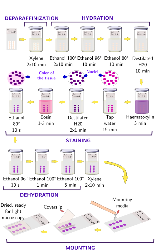
This stains information should also be considered in relation to Histology FixativesTo see related histology images use the CategoryHistology link. The process of histological staining takes five key stages which involve. Deparaffinize and hydrate to distilled water.
A special stain is a staining technique to highlight various individual tissue component once we have preliminary information from the HE stain 3. Salts - dissociate in aqueous solutions to form two ions. Peter Michalka Lucia Donárová Ústav patologickej anatómie LFUK a UN pracovisko Staré mesto Sasinkova 4 Bratislava Prof.
Current used histological stains appear to be economical quick and reliable tools for interpreting archiving and delivering essential diagnoses that could not be achieved by any other means. The process of histological staining involves five primary stages namely fixation processing embedding sectioning and staining. The process of histological staining involves five primary stages namely fixation processing embedding sectioning and staining.
A light microscope equipped with fluorescence is used to. 5Treat with Alkaline alcoholic sodium chloride working solution for 20 minutes. Gram Staining for Bacteria 7.
HE stains may stain many organisms. Fixation Fixation is the addition of special substances such as chemicals to tissues under investigation to preserve them by halting the progression of various biochemical processes that lead to degradation 1. Published September 6 2017 With the use of stains and dyes histology allows researchers to visualize particular tissue structures chemical elements within cells tissues and even microorganisms.
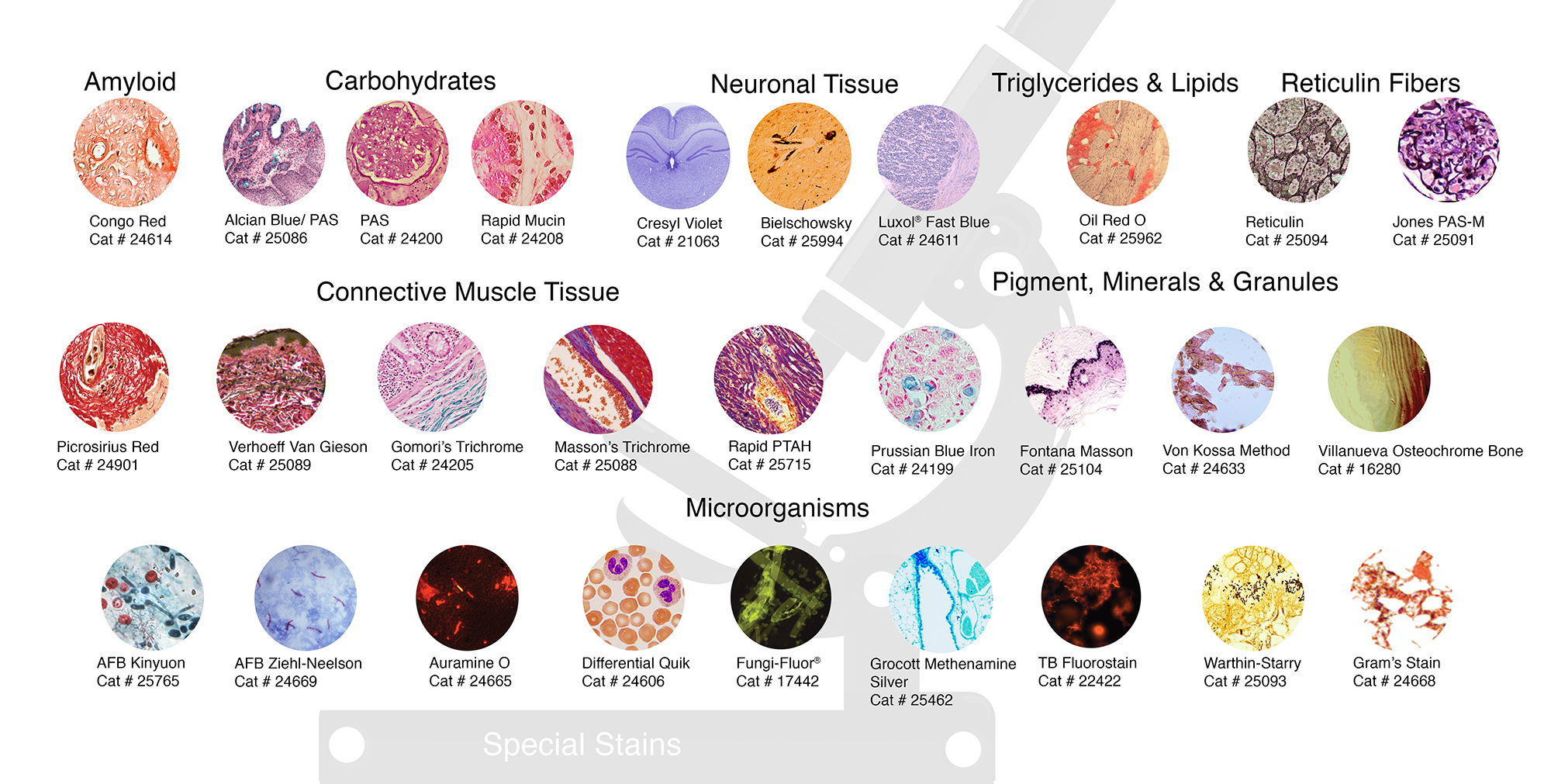
- It is the preliminary or the first stain applied to the tissue sections - Gives diagnostic information in most cases. Stains for carbohydrates 2. The advent and evolution of histology follows that of microscopy as outlined in A very Short History of Histology.
Histology stains are used to colour different. SPECIAL STAINS IN HISTOLOGY STAINS FOR MICROORGANISM CONNECTIVE TISSUE STAINS STAINS FOR PIGMENTS AND MINERAL INTRODUCTION Most infectious agents are rendered harmless by direct exposure to formal saline fixative. Blue in running water for 5minutes.
Certain stains change the coloration of cells and tissues significantly different from the color of the original dye complex a phenomenon known as metachromasia. These laboratory chemicals were potassium dichromate alcohol and the mercuric chloride to harden cellular tissues. This a specific type of stain in which primary antibodies are used that specifically label a protein and then a fluoresently labelled secondary antibody is used to bind to the primary antibody to show up where the first primary antibody has bound.
Fixation Fixation is the addition of special substances such as chemicals to tissues under investigation to preserve them by halting the progression of various biochemical processes that lead to degradation 1. Medicine Foundations students do not need to know stain information in this detail. HE stain is routine stain.
Histological staining acid basic histochemistry.

Special Stains In Histopathological Techniques Pdf Staining Dna
Staining Histology Cytology Histology Cytology Pathology Products
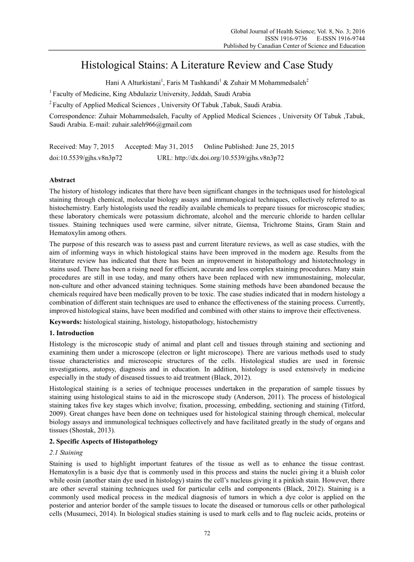
Pdf Histological Stains A Literature Review And Case Study

Pdf Computational Histological Staining And Destaining Of Prostate Core Biopsy Rgb Images With Generative Adversarial Neural Networks Semantic Scholar

Pdf Computational Histological Staining And Destaining Of Prostate Core Biopsy Rgb Images With Generative Adversarial Neural Networks Semantic Scholar
Pdf Notes On Histological Techniques
Pdf Routine Histological Techniques

Pdf A Study Of Xylene Free Hematoxylin And Eosin Staining Procedure

Lesson 12 Metachromatic Staining Pdf Staining Histology
Pdf Dyes And Stains From Molecular Structure To Histological Application

Pdf Introduction To Special Stains Semantic Scholar
Pdf Computational Histological Staining And Destaining Of Prostate Core Biopsy Rgb Images With Generative Adversarial Neural Networks Semantic Scholar

Pdf Introduction To Special Stains Semantic Scholar

Pdf Introduction To Special Stains Semantic Scholar
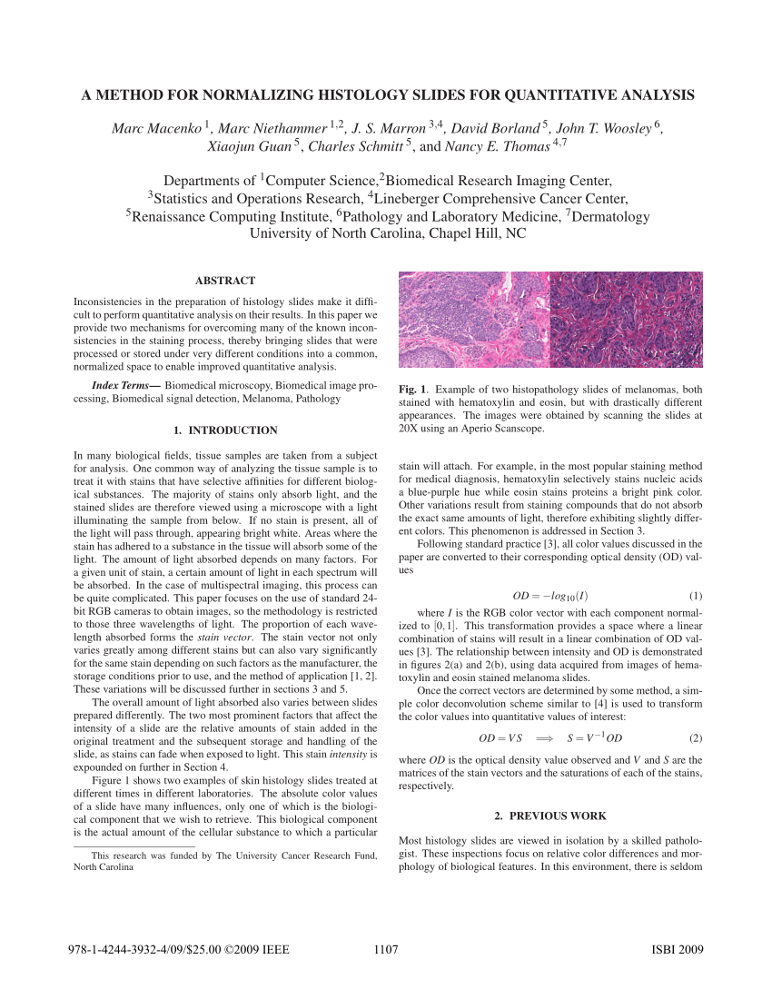
Pdf A Method For Normalizing Histology Slides For Quantitative Analysis

Special Stains Pdf Staining Histopathology

129889992 Histological Stains 2 Docx Histological Stains Aldehyde Fuchsin This Histology Stain Can Be Used To Stain Pancreatic Islet Beta Cell Course Hero
Histological Techniques 5 Staining General Staining Atlas Of Plant And Animal Histology
.jpg)
Hematoxylin Stains For Histology
0 Response to "histological stains pdf"
Post a Comment